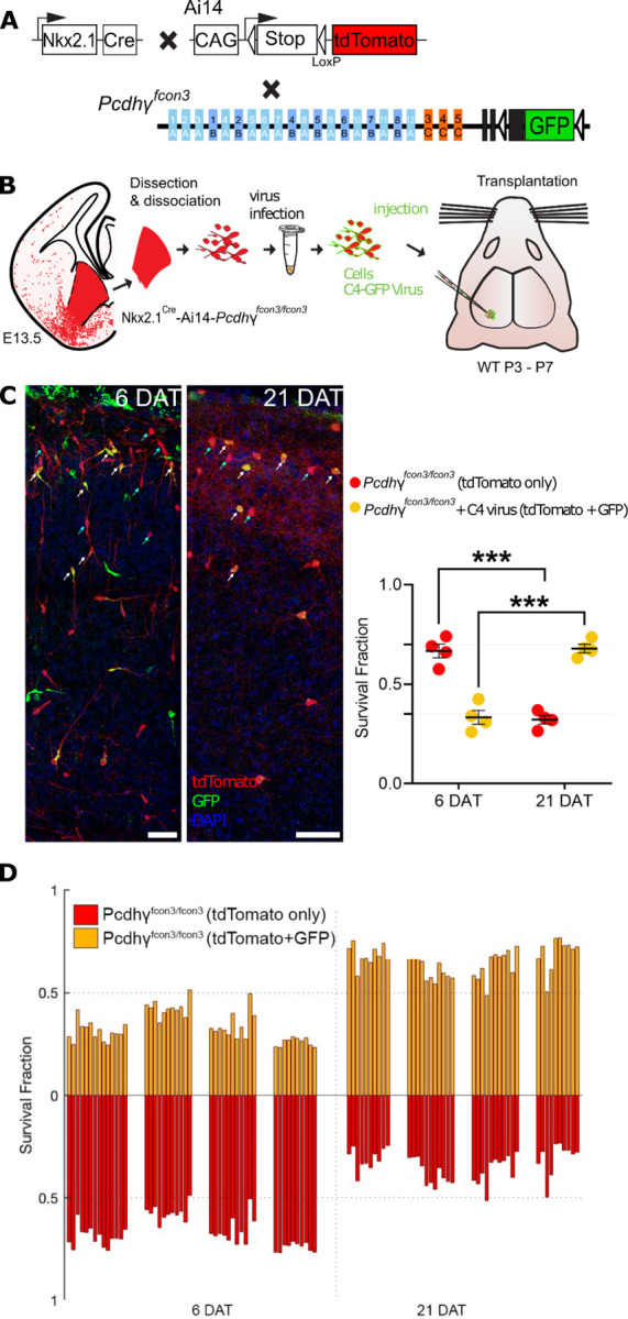
Figure 4 -. Lentiviral expression of Pcdhγc4 rescues cINs with Pcdhγ loss of function.

A. Diagram of mouse genetic crosses. Pcdhγfcon3 mice were crossed to the Nkx2.1Cre;Ai14 mouse line to generate embryos with loss of function of all Pcdhγ isoforms.
B. Schematic of the lentiviral infection and transplantation of MGE cIN precursors. The MGEs of Nkx2.1Cre;Ai14;Pcdhγfcon3/fcon3 embryos were dissected, dissociated, and infected in suspension with lentivirus carrying Pcdhγc4-GFP. The mixture of transduced and non-transduced cells was grafted into the cortex of WT neonate recipient mice.
C. Confocal images of the transplanted cINs in the cortex at 6 and 21 DAT. Notice the expression of GFP can be found near the cell surface including the cell processes, reflecting the putative location of the transduced Pcdhγc4-GFP protein. Quantifications of the tdTomato+GFP-(teal arrows) or tdTomato+GFP+ (yellow cells, white arrows) cells are shown as the fraction of cells from the total tdTomato+ cells at 6 and 21 DAT. The fraction of the Pcdhγc4-transduced yellow cells increases from 6 to 21DAT, while the fraction of non-transduced (tdTomato+ GFP-) decreases at equivalent time points.
D. Survival fraction quantification from (C) shown by the brain section (each bar) and separated by animals at 6 and 21 DAT.
Scale bar = 50 um, Nested-ANOVA, ****p = 0.0002, n = 4 mice per time point and 10 brain sections per mouse from one transplant cohort.
