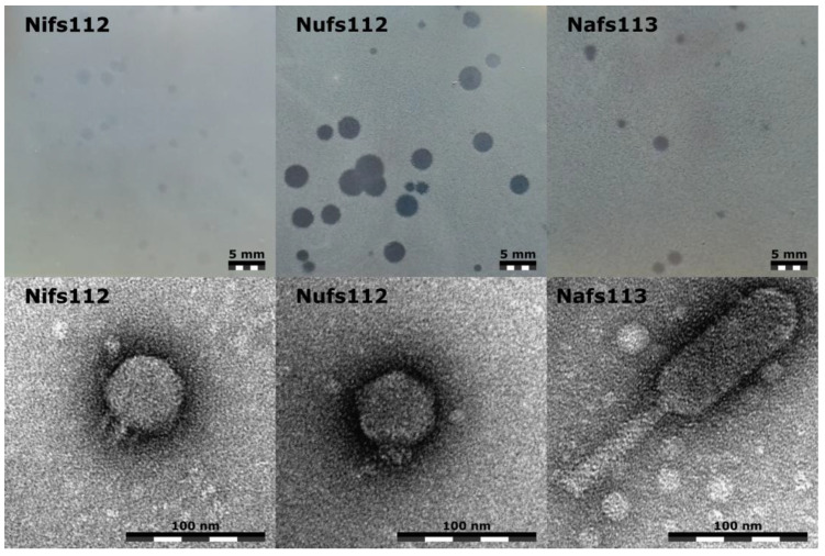
Figure 2.
Plaque (upper row) and virion morphologies (lower row) of the studied Pantoea phages Nifs112, Nufs112, and Nafs113. Plaques were photographed after incubation for ~24 h at room temperature using P. agglomerans LS5-2 culture for the lawn and the same double agar overlay plating conditions; the scale bar represents 5 mm. Representative virion micrographs were obtained from the corresponding phage purified lysates negatively stained with 0.5% uranyl acetate; the scale bar represents 100 nm.

