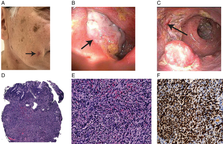
Figure 1.
(A) Skin lesion (arrow) on the right cheek with biopsies returning positive for Kaposi sarcoma. (B) Large ulcer (arrow) (>2-cm) deeply cratered ulcer noted in the sigmoid colon, biopsy-positive for Kaposi sarcoma. (C) Large (>2-cm) deeply cratered ulcer (arrow) noted in rectum, biopsy-positive for Kaposi sarcoma. (D) Low-power view shows an infiltrative spindle cell proliferation occupies the colonic mucosa suggestive of KS (H&E 40×, rectal ulcer biopsy). (E) Higher-power view shows the tumor cells with uniform spindled nuclei and extensive red blood cell extravasation, suggestive of KS (H&E 200×, rectal ulcer biopsy). (F) IHC stain for human herpesvirus-8 shows diffusely and strongly nuclear staining in the spindled tumor cells, suggestive of KS (IHC, 200×, rectal ulcer biopsy). H&E, hematoxylin and eosin; IHC, immunohistochemical; KS, Kaposi sarcoma.

