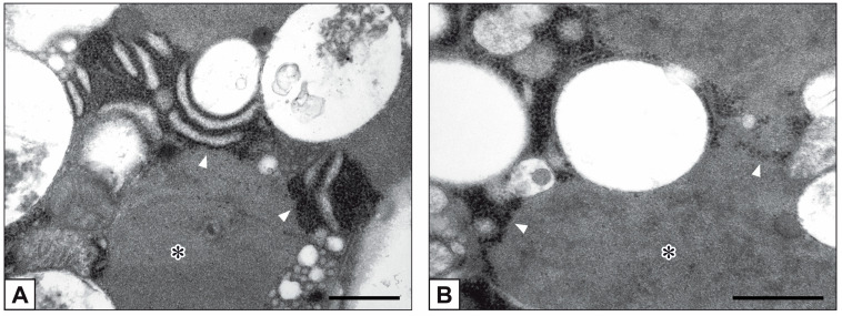
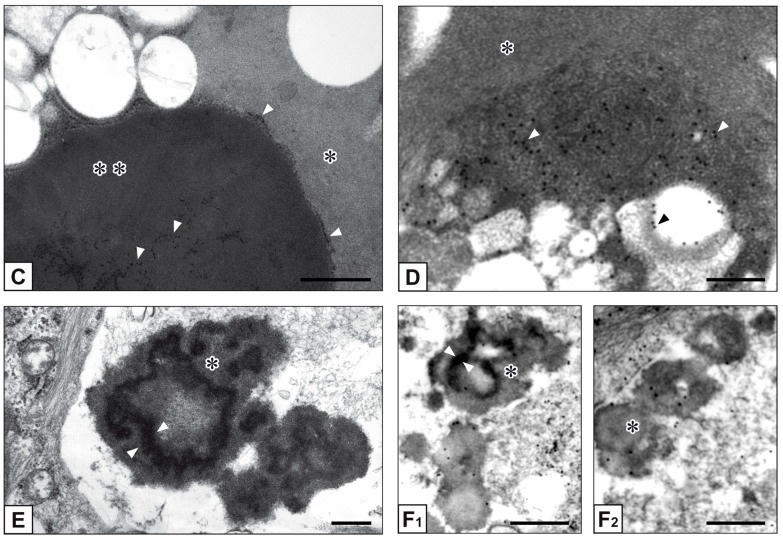
Figure 2.
Ultrastructure of bAVICs from 15-day-long pro-calcific cultures. (A–C) Enlargement of RER cisternae (arrowheads) and RER membrane lysis, with ribosome release and merging with the nearby phthalocyanine-positive material (single asterisk: less condensed PPM; double asterisk: more condensed PPM) derived from ongoing membranous organelle degeneration. (D) Immunogold labelling of rRNA with gold particles (white arrowheads) decorating the intracellular PPM (asterisk) and a cross-sectioned degenerating RER cisterna (black arrowhead). (E) Cell-derived byproducts showing a mix of PPM (asterisk) and more electrondense phthalocyanine-positive layers (PPLs; counterposed arrowheads). (F1,F2) Immunogold labelling of rRNA showing gold particles within PPM (asterisks) and PPLs (counterposed arrowheads) at the level of vesicular cell debris. Bar: 0.5 μm (A–C,E); 0.25 μm (D,F1,F2).


