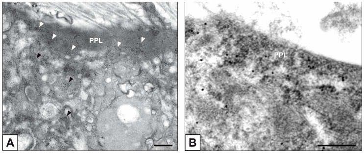
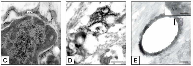
Figure 6.
Ultrastructure of AVICs populating stenotic hAVLs. (A) Ribosomes (black/white arrowheads) embedded within PPM/PPL in a mineralizing hAVIC. (B,C) Immunogold labelling of rRNA with gold particles decorating peripheral PPLs. (D) Immunogold labelling of rRNA showing gold particles within PPM/PPLs in a hAVIC undergone advanced calcific degeneration. (E) Immunogold labelling of rRNA showing gold particles onto a PPL lining a vesicle-shaped cell byproduct. Inset: magnification of the PPL-squared region. Bar: 0.25 μm (A–E); 0.1 μm ((E) inset.


