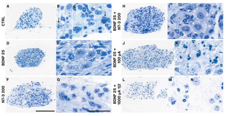
Figure 9.
Semithin sections of BDNF and/or NT-3 supplemented, and electrical stimulated explant cultures. Treatments influenced the cell morphology of SGNs (black stars) and ensheathing SGCs (white stars). Control explants without neurotrophic supplementation (A–C) exposed many degenerating neurons (arrows) and several SGCs. Explants after 25 ng/mL BDNF treatment (D,E) resembled a normal morphology with many SGCs ensheathing SGNs with big nucleoli. 200 ng/mL NT-3 administration (F,G) resulted in big neurons but fewer SGCs associated with these neurons. Combined BDNF and NT-3 (H,I) application showed a similar appearance as with BDNF alone, while ES at 100 µA (J,K) and 1000 µA and a 1 min on, 2 min off pattern (L–N) lead to a marked decline of SGCs. Often SGNs located without their SGCs in the explant tissue (L,N). Scale bars: 100 µm (overview) and 10 µm (detail).

