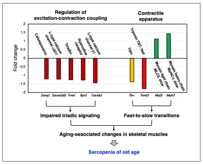
Figure 3.
Representative example of the mass-spectrometry-based proteomic analysis of skeletal muscle aging. Shown are crucial regulatory proteins of excitation–contraction coupling and Ca2+- homeostasis (dihydropyridine receptor L-type Ca2+-channel, ryanodine receptor Ca2+-release channel, calsequestrin and triadin) and sarcomeric proteins (titin, troponin, myosin light chain and myosin heavy chain). Mass spectrometric analyses of young versus aged wild-type mouse diaphragm muscle specimens were carried out as previously described in detail [201,269,333].

