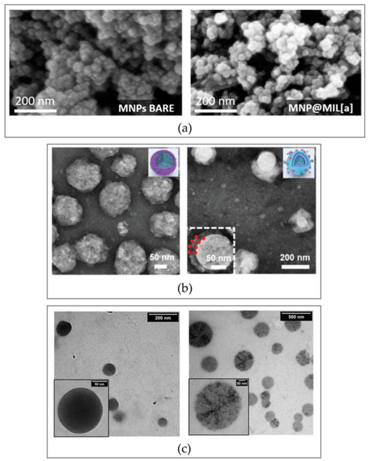
Figure 3.
Morphological characterization of surface-modified NPs. (a) Fe3O4 magnetic NPs covered with a metal–organic framework. SEM images of bare (left) and decorated (right) NPs. Adapted with permission from Pulvirenti et al. [143] (IJMS, MDPI, 2022). (b) TEM analysis of biomimetic NPs made up of siRNA complexed polyethyleneimine xanthate before (left) and after the cell membrane coating (right). Adapted with permission from Zhang et al. [154] (Adv. Funct. Mater., Wiley, 2022). (c) TEM images of hyaluronic acid NPs pre (left) and post (right) modification with Angiopep-2. Adapted with permission from Costagliola di Polidoro et al. [155] (Cancers, MDPI, 2021). Abbreviations: NPs—nanoparticles; SEM—scanning electron microscopy; TEM—transmission electron microscopy.

