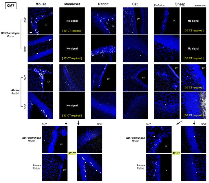
Figure 6.
Representative confocal images of Ki67 antigen detection (in white) in different mammals for two different antibodies in the two neurogenic sites (forebrain SVZ and hippocampal SGZ). All specimens are counterstained with DAPI. All photographs have been performed at the same magnification (scale bar: 30 µm). In marmoset, (30′ CT) indicates that the antigen can be detected only after 30 min of citrate buffer treatment. LV: lateral ventricle.

