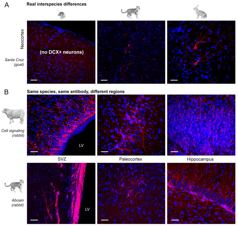
Figure 7.
Variation in the type and quality of immunostaining obtained in different animal species (A) and in different brain regions (B). (A) In comparative studies the detection DCX+ cell populations can yield different results depending on real interspecies differences, e.g., the absence of DCX+, immature neurons in the mouse neocortex [23]. (B) Even in the same species, using the same antibody, different results can be found depending on the brain region. In most cases, the SVZ bordering the lateral ventricle (LV) stains far more clean and clear with respect to parenchymal regions such as the cortex and the hippocampus (sheep and cat). Note that in sheep, SVZ and cortex stain for the expected neuronal populations and non-specific staining is detectable in the hippocampus. Scale bar: 30 µm.

