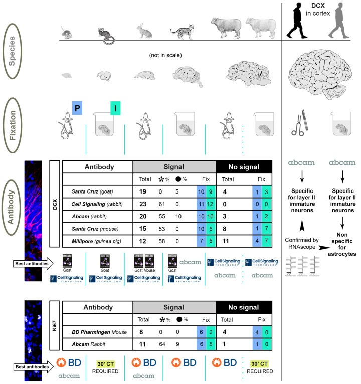
Figure 10.
Schematic summary of the main results and conclusions. The three main variables considered in the study are reported in grey ovals on the left, including the animal species (characterized by widely different brain sizes), the type of fixation (perfusion: P, blue squares; immersion: I, green squares), and the commercial antibodies tested for DCX and Ki67 antigen. In the tables, the numbers of staining samples are reported (analysed in the four brain regions of the five mammalian species, each corresponding to two cryostat sections from three animals, treated with 5′ citrate), for a total of 115 staining samples for DCX (120 including the mouse neocortex lacking DCX+ cells), and 24 for Ki67. On the whole, a total of 89 staining samples for DCX made a positive signal, while 26 did not. For Ki67, there were 19 with positive staining, and 5 negative. The “No signal” column was considered to also include non-specific staining (i.e., non-successful staining). It is worth noting that the number of specimens fixed with perfusion or immersion (reported in blue and green squares, respectively) showing successful staining were almost equally distributed, especially for DCX. In the case of Ki67, we showed that some specimens fixed by immersion require longer time of citrate treatment to reveal staining (see Figure 6). The best-performing antibodies for each animal species are reported in tables below. Also in this case, the result is mostly independent from fixation (see for example the same outcome for DCX staining in perfused and immersed sheep brains), and rather linked to the association between animal species and antigen considered. In humans, only the Abcam antibody delivered a satisfactory result; on intraoperative specimens, the specificity of the staining was confirmed by RNAscope analysis (see Figure 9), also revealing a non-specific staining on astrocytes in the post-mortem tissue. Logos reproduced with authorization of Companies.

