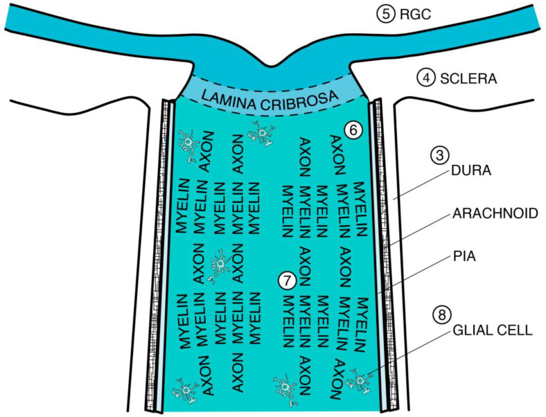
Figure 1.
Schematic anatomical image of the optic nerve pertaining to age-dependent neurodegeneration. Numbers correspond with section headings of the present review, specifically “3. Connective Tissue,” which includes “3.4. Dura Mater,” “3.5. Arachnoid and Subarachnoid Space,” “3.6. Pia Mater and Septa,” and “3.7. Lamina Cribrosa (LC)”; “4. Decreased Optic Canal Expansion”; “5. Retinal Ganglion Cell (RGC)”; “6. Axon”; “7. Myelin”; and “8. Glial Cell.”

