Figure 4.
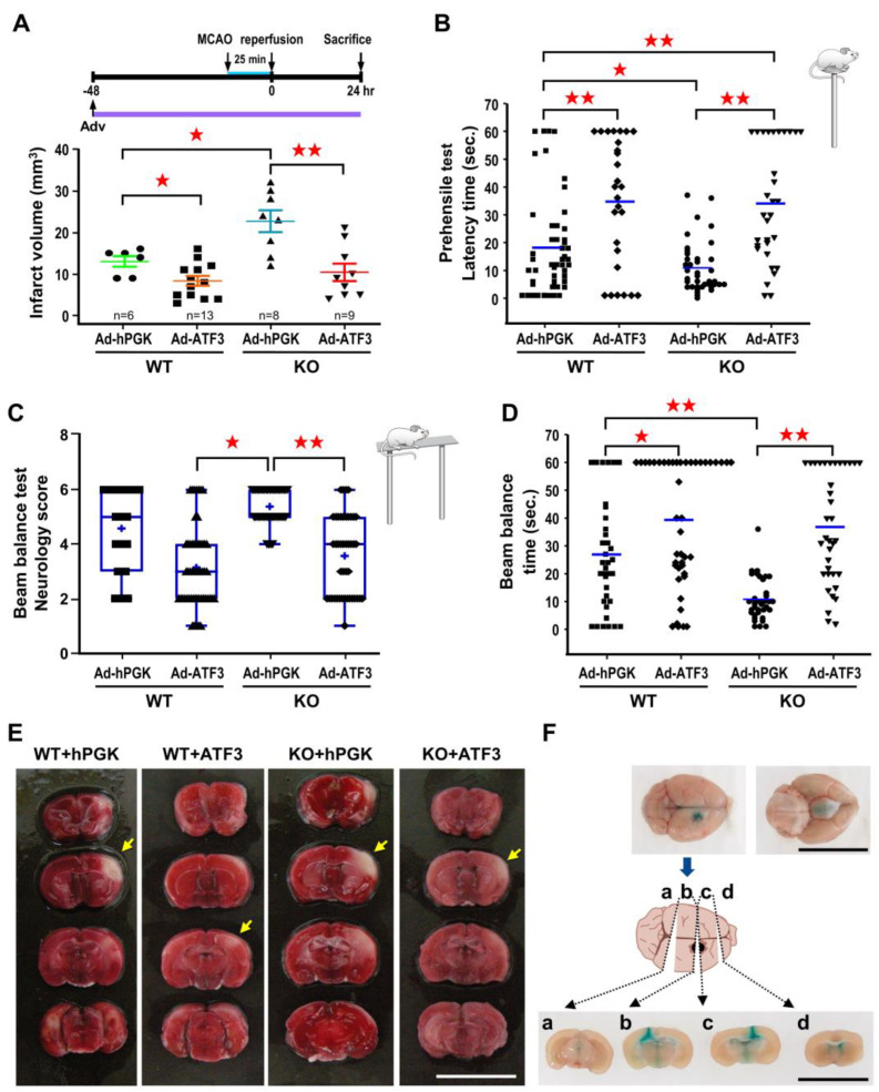
ATF3 overexpression rescues brain infarct and neurological deficits in ATF3 KO mice. ATF3 KO or WT mice were subjected to icv infusion of 2 × 10⁵ pfu of Ad-ATF3 or Ad-hPGK at 48 h prior to 25 min of ischemia and infract volume was determined at 24 h of reperfusion. Brain infarct volumes are quantified in panel (A). Each dot represents a data point from an individual animal. Behavior tests based on prehensile test (B) or beam balance test (C,D) were performed at 24 h of reperfusion. Data are expressed as mean ± SD (n ≧ 6). ★ p < 0.05 and ★★ p < 0.01 between groups. Representative images of brain infarct are shown in panel (E); white area represents infarct area (yellow arrow). Periodic confirmation of proper placement of the needle was performed with infusion of fast green (F). (Bars = 1 cm). ATF3: activating transcription factor 3; Ad-hPGK: rAd-carrying hPGK promoter; hPGK: human phosphoglycerate kinase; rAd: replication-defective recombinant adenoviral; Ad-ATF3: rAd-carrying hPGK promoter-driven ATF3 gene; MCAO: middle cerebral artery occlusion.

