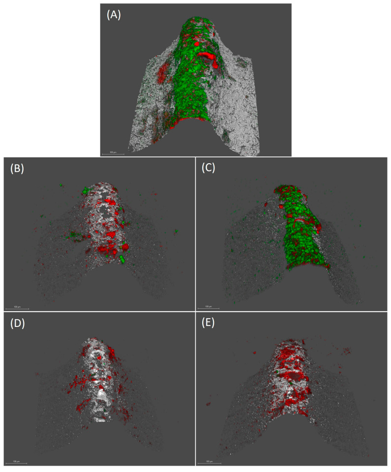
Figure 3.
Images obtained by confocal laser scanning microscopy (CLSM) at 72 h over implants treated with phosphate buffer saline (PBS) (A), 0.2% chlorhexidine (CHX) (B), 2.5% dimethyl sulfoxide (DMSO) (C), µM xanthohumol 100 (D) and 5 mM curcumin (E) (scale bar = 100 µm). LIVE/DEAD® BackLight Kit was used. Live bacteria (green), dead bacteria (red) and implant surface (white) can be differentiated.

