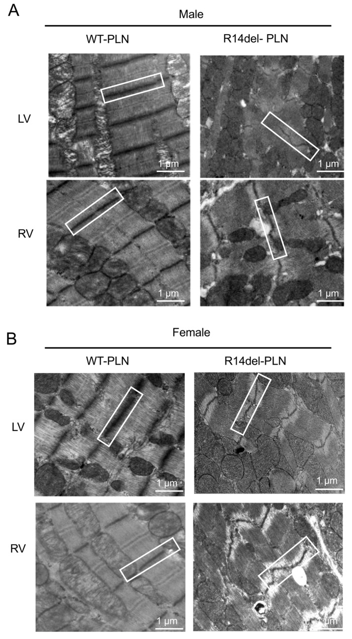
Figure 8.
Transmission electron microscopy (TEM) evaluations of left ventricle (LV top panels) and right ventricle (RV bottom panels) from 12-month-old male (A) and female (B) WT-PLN and R14del-PLN mice. Representative images showed irregular/disarrayed, widened Z-disc patterns as well as streaming and smearing of the Z-disc material in R14del ventricles (white box). N = 4 per group. Scale bars: 1 μm, magnification 6000×.

