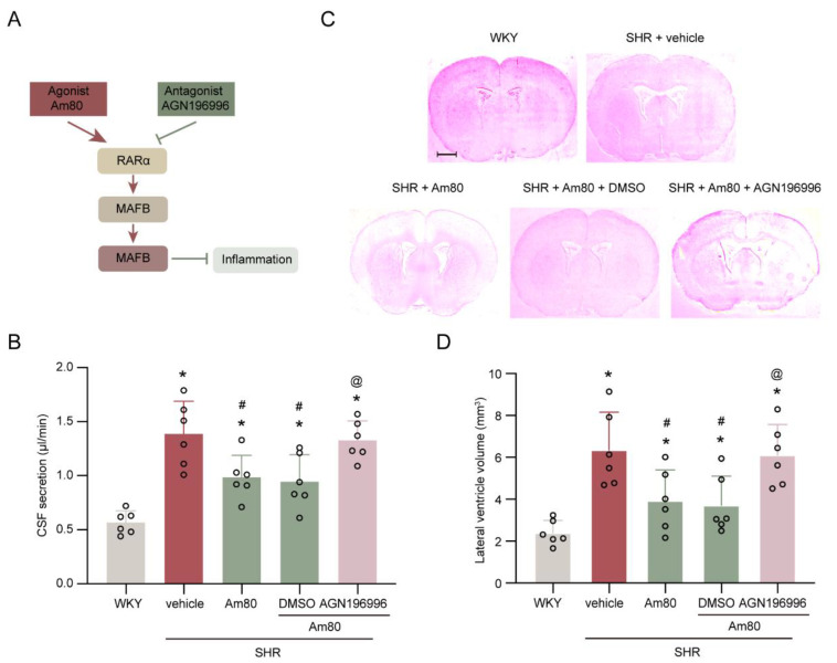
Figure 3.
AGN196996 abolished the protective effects of Am80. (A) Schematic diagram of the experimental design. (B) Quantification of the rate of CSF secretion at week 7 (n = 6). (C) Representative photomicrographs of coronal sections of rat brains (at −0.6 mm from the bregma), depicting ventricular volume at week 7. Scale bar, 2 mm. (D) Quantification of lateral ventricle volume at week 7 (n = 6). * p < 0.05 versus WKY rats at week 7, # p < 0.05 vs. SHR + vehicle group, @ p < 0.05 vs. SHR + Am80 + DMSO group. Values are means ± SD.

