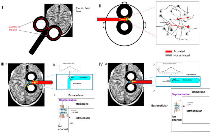
Figure 2.
(I). Electric field lines induced by a typical figure-8 TMS coil. (II). Under the figure-8 central segment, only neurons aligned parallel to the electric field, along the coil axis, are activated (indicated in red). (III). Where neurons under the coil center have axon parallel to the induced electric field and bends away from it (a), the field is maximal at the bend point (b) and leads to transmembrane potential Vm across the voltage-gated ion channels (c), which are then opened, and action potential is initiated. (IV). Where neurons under the coil center have axon parallel to the induced electric field and terminate (a), the field is maximal at the axon terminal (b) and leads to transmembrane potential Vm across the voltage-gated ion channels (c), which are then opened, and action potential is initiated.

