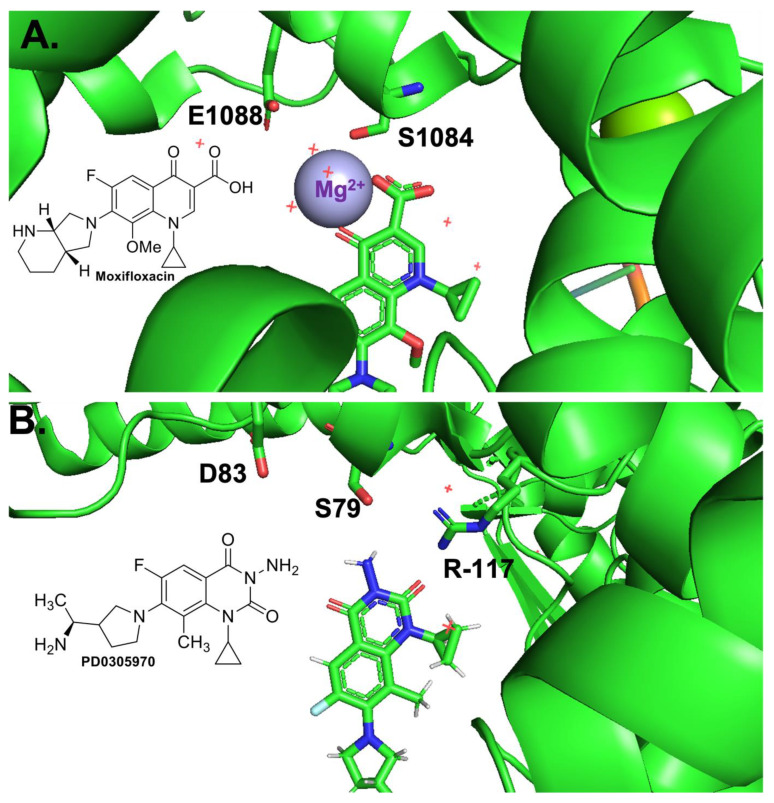
Figure 1.
Crystal structures of a quinolone and a quinazolinedione interacting with topoisomerase IV-DNA cleavage complexes. (A) Crystal structure of moxifloxacin, DNA, and A. baumannii topoisomerase IV. Fluoroquinolone and key residues displayed as sticks, water molecules displayed as red +, divalent magnesium ion displayed in lavender. Adapted from RCSB PDB: 2XKK, visualized with The PyMOL Molecular Graphics System, Version 2.5.2 Schrödinger, LLC. Chemical structure of Moxifloxacin is shown in the inset. (B) Crystal structure of PD0305970, DNA, and S. pneumoniae topoisomerase IV. Quinazoline-2,4-dione displayed as sticks. Adapted from RCSB PDB: 3RAF, visualized with The PyMOL Molecular Graphics System, Version 2.5.2 Schrödinger, LLC. Chemical structure of PD0305970 is shown in the inset.

