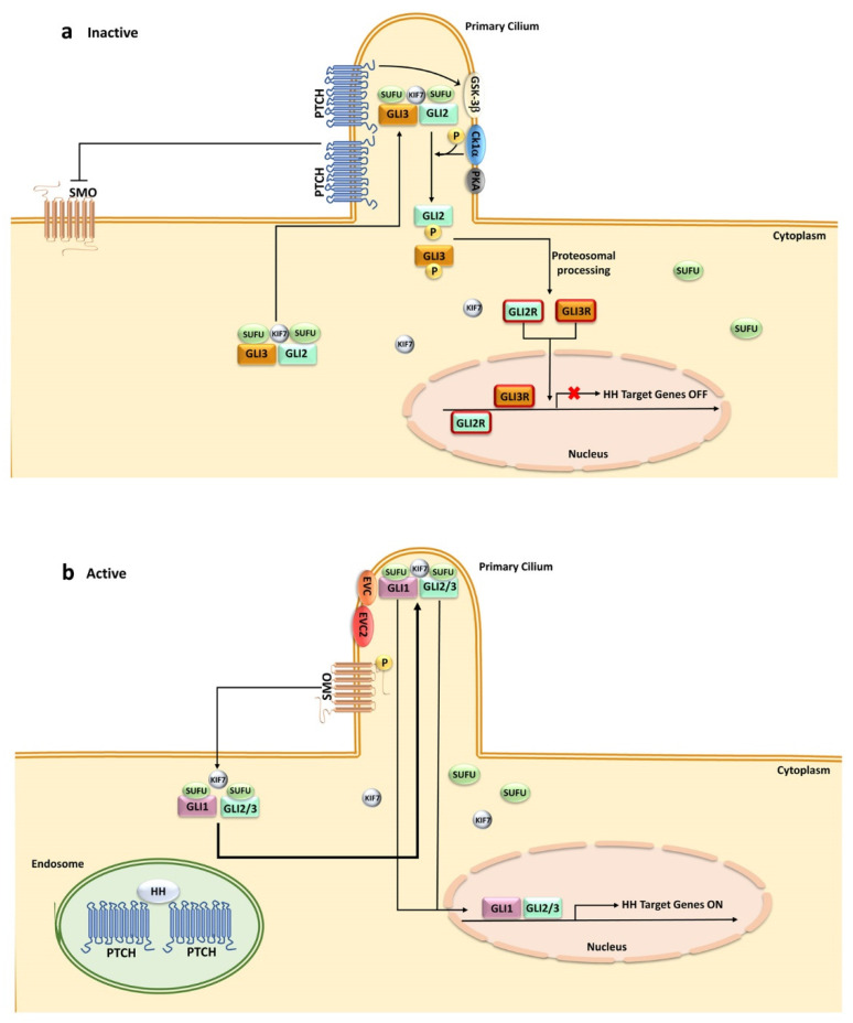
Figure 1.
HH signaling in mammals. A simplified cartoon illustrating activation of HH pathway in mammalian cells: (a) in the absence of HH ligands, PTCH is localized at the primary cilium while SMO remains distant from the cilium. PTCH inhibits SMO by preventing its cilial entry. Several kinases (GSK-3β, CK1-α, PKA) are recruited by the GLI/SUFU/KIF7 complexes and activated by PTCH. GLI transcription factors are phosphorylated by these kinases, which promote their processing into the repressor forms, translocating to the nucleus, and thereby blocking HH target gene transcription; (b) HH ligand binding to PTCH relieves SMO inhibition. PTCH is endocytosed and displaced from the cilium, whereas SMO accumulates in the cilium in an active state. The GLI/SUFU/KIF7 complexes are recruited by SMO. SMO activation results in the detachment of GLI transcription factors from SUFU and KIF7 with the aid of EvC and EvC2. GLI transcription factors migrate to the nucleus, activating gene transcription. CK1-α, casein kinase 1-α; EvC, Ellis–Van Creveld Syndrome; GLI-R, GLI repressor; GSK-3β, glycogen synthase kinase 3β; KIF7, kinesin family protein 7; PKA, protein kinase A; PTCH, Patched; SMO, Smoothened; SUFU, suppressor of fused.

