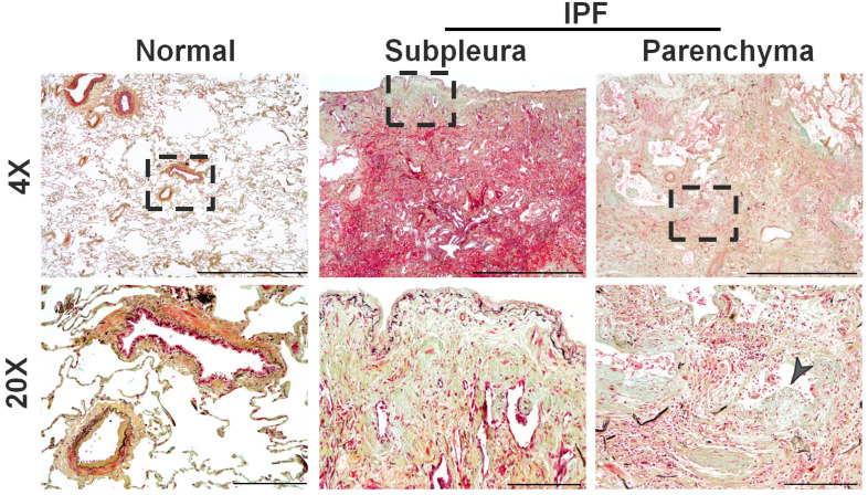
Figure 1.
Representative images of Movat Pentachrome-stained distal areas of normal and IPF lungs. Highlighted dashed region in low magnification (Scale bar, 1500 µm) images represents the high magnification (Scale bar, 200 µm) images that show the prominent subpleural thickening and fibrotic foci that accumulate in the distal areas of the alveolar parenchyma of IPF lungs compared to normal lungs. Pentachrome staining highlights collagen (yellow color), muscle (red color), and elastic fibers (black to blue color) in mature fibrotic lesions of IPF. Arrowhead is used to highlight the fibrotic foci.

