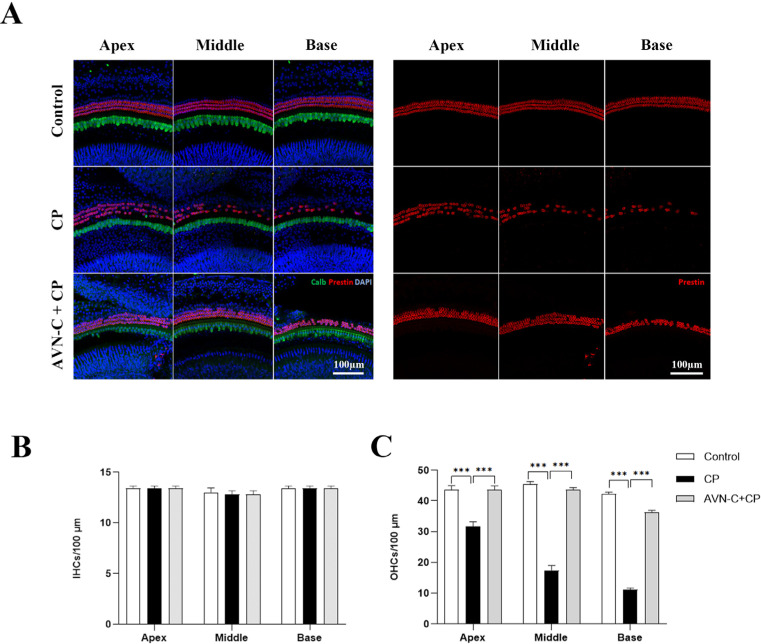
Figure 2.
Immunohistochemical staining of the organ of Corti. (A) Right panel: images of OHCs stained with Prestin (red); left panel: combined image of Calbindin (green) for IHCs, Prestin for OHCs, and DAPI (blue) staining. (B) The IHCs remained unchanged in all three groups (n = 5 each). (C) The OHCs were damaged severely by CP treatment (32 ± 3.4, 17 ± 3.4, and 11 ± 1.4 OHCs per 100 µm at apex, middle, and base turns, respectively) whereas AVN-C protected OHCs from the CP-induced loss (44 ± 1.5, 44 ± 1.7, and 36 ± 2.9 OHCs per 100 µm at the apex, middle, and base turns, respectively). The OHCs in the control group were found at the apex (45 ± 3.0), middle, (44 ± 2.1), and base turns (42 ± 1.5 OHCs) per 100 µm, respectively. (*** p ≤ 0.001, p ≤ 0.05 was considered significant in all experimental groups).

