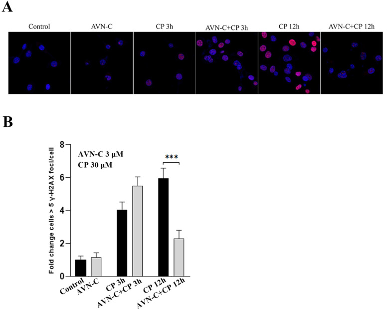
Figure 6.
AVN-C prevents DNA damage caused by CP. AVN-C 3 µM and CP 30 µM were used in this study. (A) Representative images of γH2AX-stained HEI-OC1 cell. (B) γH2AX levels in relation to exposure to CP and AVN-C at different timepoints was as follows: Control (1 ± 0.2), AVN-C (1.2 ± 0.3), CP 3 h (4.1 ± 0.5), AVN-C+CP 3 h (5.5 ± 0.6), CP 12 h (6.1 ± 0.6), and AVN-C+CP (2.3 ± 0.5) fold-change cells > 5 γH2AX foci/cell and (*** p ≤ 0.001, CP vs AVN-C. + CP-treated groups at 12 h). One-way ANOVA was used and p ≤ 0.05 was considered significant. Image magnification 40X was used.

