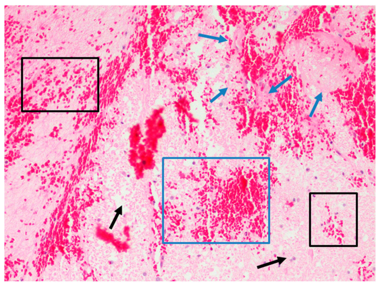
Figure 2.
H&E section (×20) showing distribution pattern of erythrocyte extravasations in cerebral ischemia. Intraparenchymal zone in which erythrocyte extravasations are partly diffused (black box), partly wider (blue box), in a context of ischemic necrosis (blue arrow) and edema with aspects of hypoxic cell damage (black arrow).

