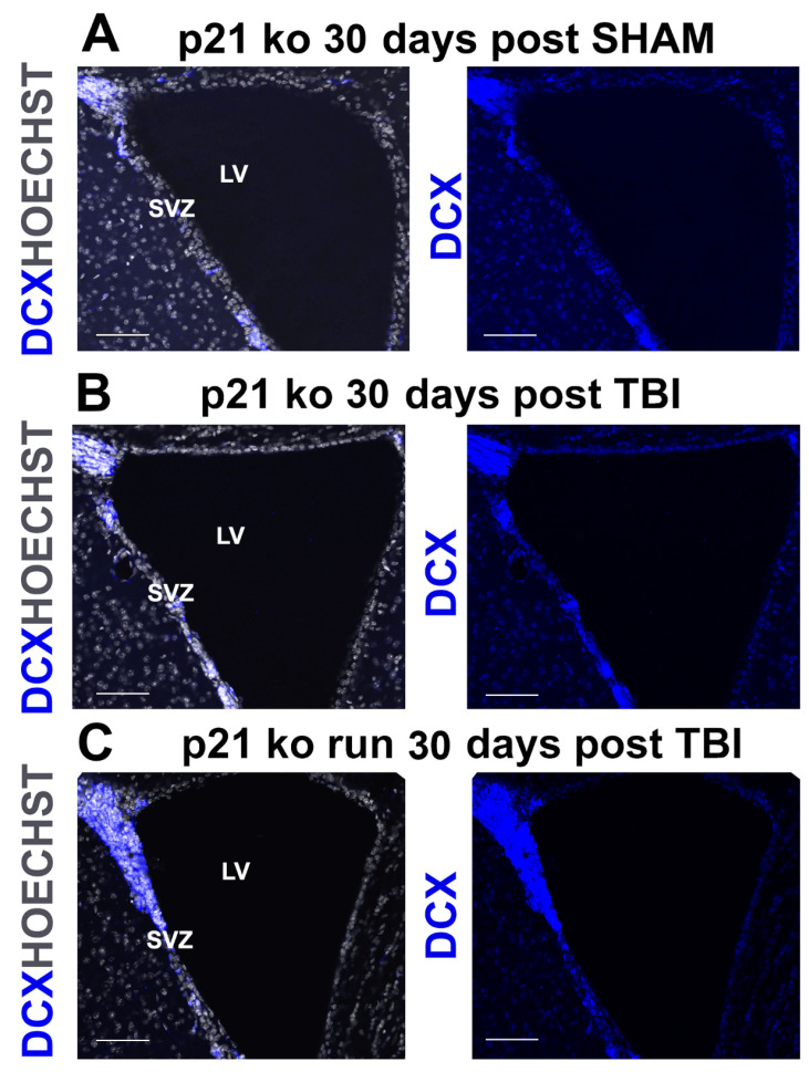
Figure 4.
(A–C). Increase in DCX+ cells in the KO RUN TBI group. Confocal representative micrographs show the increased number of DCX+ cells in the KO RUN TBI mice (C), in comparison with the KO SHAM (A) and KO TBI (B) mice, 30 days after TBI. Magnification = 20×. Scale bar = 100 μm. SVZ = subventricular zone. LV = lateral ventricle.

