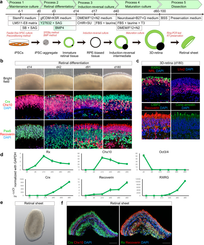
Fig. 1. Self-organizing culture of human iPSCs to generate the 3D-retina and dissected retinal sheet.
a Scheme of the self-organizing culture. b Bright-field view of iPSC-S17-derived cell aggregate containing retinal tissue on days 14, 42, 91, and 180 (upper). Scale bar in bright-field view: 100 µm. Immunostaining of iPSC-S17-derived retinal tissue on days 14, 42, 91, and 180 (middle and lower). Crx (green) and Chx10 (red) in middle panels. Pax6 (green) and Recoverin (red) in lower panels. Blue: nuclear staining with DAPI. Scale bar in immunostaining: 20 µm. c Immunostaining of iPSC-S17-derived retinal tissue on day 180. RXRG (green) and NRL (red) in upper-left panel. Lhx2 (red) in upper-right panel. CtBP2 (green) and Recoverin (red) in lower-left panel. Cone-arrestin (green) in lower-right panel. Blue: nuclear staining with DAPI. Scale bar: 20 µm. d Gene expression analysis in iPSC-S17-derived cell aggregates containing retinal tissue on days 0, 6, 14, 45, 60, and 80. In each replicate, RNA was extracted from 48 aggregates. mRNA levels were determined by qPCR analysis. Relative mRNA expression was determined by the delta-delta Ct method with GAPDH as an endogenous control. Data are presented as mean ± SE (n = 4 per time point). e Bright-field view of iPSC-S17-derived retinal sheet. Scale bar: 100 µm. f Immunostaining of iPSC-S17-derived retinal sheet on day 87. Crx (green) and Chx10 (red) in left panel. Rx (green) and Recoverin (red) in right panel. Blue: nuclear staining with DAPI. Scale bar: 100 µm.

