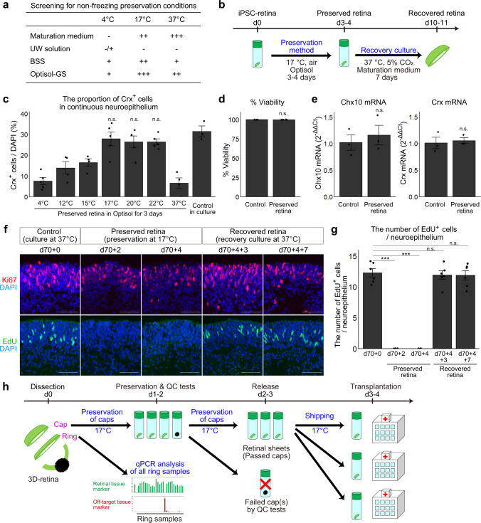
Fig. 4. Controlled room temperature non-freezing preservation method.
a Screening for the non-freezing preservation conditions. +++: multilayered continuous retinal epithelium was preserved. ++: multilayered continuous retinal epithelium was preserved but neural rosettes appeared. +: multilayered continuous retinal epithelium was observed but very limited. –, no multilayered continuous retinal epithelium was observed after the preservation. b Scheme of the preservation experiments. c Proportions of Crx+ photoreceptor precursors in multilayered retinal tissues. The proportions were calculated by dividing the number of Crx+ cells by the number of nuclei (DAPI). Data are presented as mean ± SE for n = 5 (4 °C), 4 (12 °C), 4 (15 °C), 5 (17 °C), 5 (20 °C), 5 (22 °C), 4 (37 °C), and 3 (control culture). d Cell viability in non-preserved retinas (Control) and retinas preserved for 4 days. Data are shown as mean ± SE (n = 5 per group). e Gene expressions of Chx10 and Crx in non-preserved retinas (Control) and retinas preserved for 4 days. Data are presented as mean ± SE (n = 3 per group). f IHC analysis of retinas that were not preserved (d70 + 0), preserved in Optisol for 2 days (d70 + 2) and 4 days (d70 + 4), and preserved in Optisol for 4 days followed by recovery culture for 3 days (d70 + 4 + 3) and 7 days (d70 + 4 + 7). Ki67 (red) staining in the upper panels. EdU staining (green) in lower panels. DAPI is shown in blue. Scale bar: 50 µm. g Numbers of EdU-positive cells per 100 µm-wide multilayered retinal tissue in each group. Data are presented as mean ± SE (n = 6 per group). ***p < 0.001 for one-way ANOVA followed by a Tukey’s test. h Scheme for dissection, ring-PCR test, and shipping of retinal sheets. n.s., not significant.

