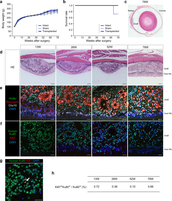
Fig. 5. In vivo tumorigenicity study by subretinal transplantation in nude rats.
Time courses of body weight (a) and Kaplan–Meier survival curves (b) in nude rats in the tumorigenicity study. Intact: control nude rat without surgery (n = 4). Sham: control nude rat with subretinal injection of vehicle control (n = 4). Transplanted: nude rat with subretinal transplantation of a single iPSC-Q-derived retinal sheet (n = 4). Data are presented as mean ± SE. c, d HE staining of transplanted eye sections at 13, 26, 52, and 78 weeks after transplantation. Scale bar: 100 µm. e–h Immunostaining of transplanted eye sections for human nuclear markers, retinal markers, and Ki67. Scale bar: 20 µm. e Immunostaining for HuNu (green), Recoverin (red), Chx10 (white), and DAPI (blue). f Immunostaining for human Ku80 (green), Ki67 (red), and DAPI (blue). g High-magnification image of immunostaining for the graft (52 weeks) with antibodies for human Ku80 (green), Ki67 (red), and DAPI (blue). Arrow indicates Ki67+ and Ku80+ cells. h Percentages of Ki67+ and Ku80+ cells among the Ku80+ human cells. Data are presented as mean. INL inner nuclear layer.

