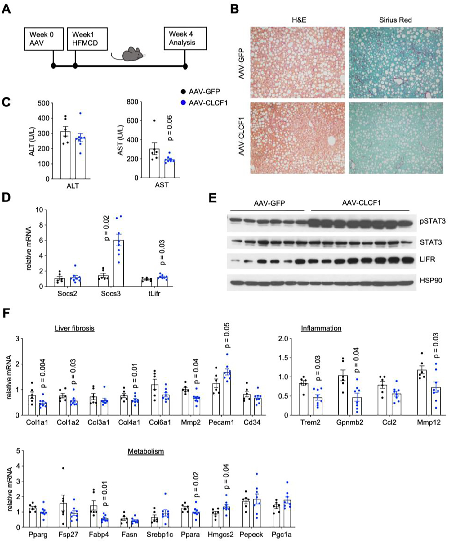
Figure 5. AAV-mediated CLCF1 overexpression in the liver ameliorates NASH pathologies in mice fed CDA-HFD diet.

A) A schematic outline of study design.
B) Liver histology from transduced mice (H&E, left; Sirius Red staining, right).
C) Plasma ALT and AST levels in mice transduced with AAV-GFP (n=6) or AAV-CLCF1 (n=7) after six weeks of CDA-HFD diet feeding.
D) qPCR analysis of hepatic genes involved in IL-6 family cytokine signaling.
E) Immunoblots of liver lysates from transduced mice fed with CDA-HFD diet for six weeks.
F) qPCR analysis of genes involved in liver fibrosis, vascular endothelium, inflammation, and hepatic metabolism in transduced mice.
Data in C), D) and F) represent mean ± SEM; two-tailed unpaired Student’s t-test.
