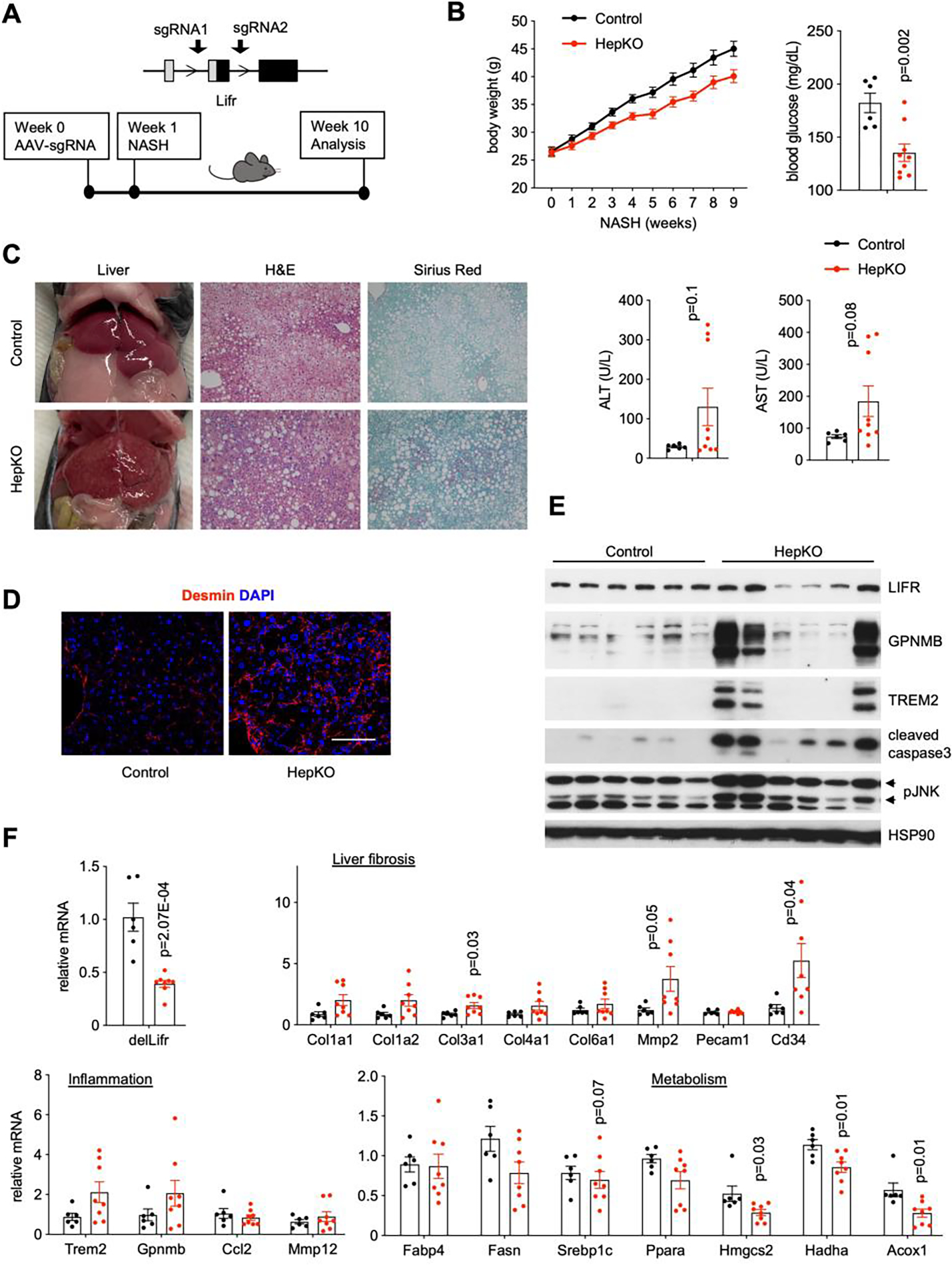
Figure 6. Hepatocyte-specific deletion of LIFR accelerates NASH progression.

A) A diagram of Cas9-mediated Lifr deletion and study design. Albumin Cre negative/Rosa26-FSF-Cas9 (control) and Cre+/Rosa26-FSF-Cas9 (HepKO) littermates were transduced with AAV-sgRNAs targeting Lifr via tail vein injection. Transduced mice were subjected to NASH diet feeding for nine weeks.
B) Metabolic and NASH parameters of control (n=6) and HepKO (n=8) mice.
C) Liver appearance and histology in control and HepKO mice following ten weeks of NASH diet feeding.
D) Desmin immunofluorescence staining. Scale bar = 100μm.
E) Immunoblots of liver lysates from control and HepKO mice fed NASH diet.
F) qPCR analysis of hepatic gene expression.
Data in B) and F) represent mean ± SEM; two-tailed unpaired Student’s t-test.
