FIGURE 3.

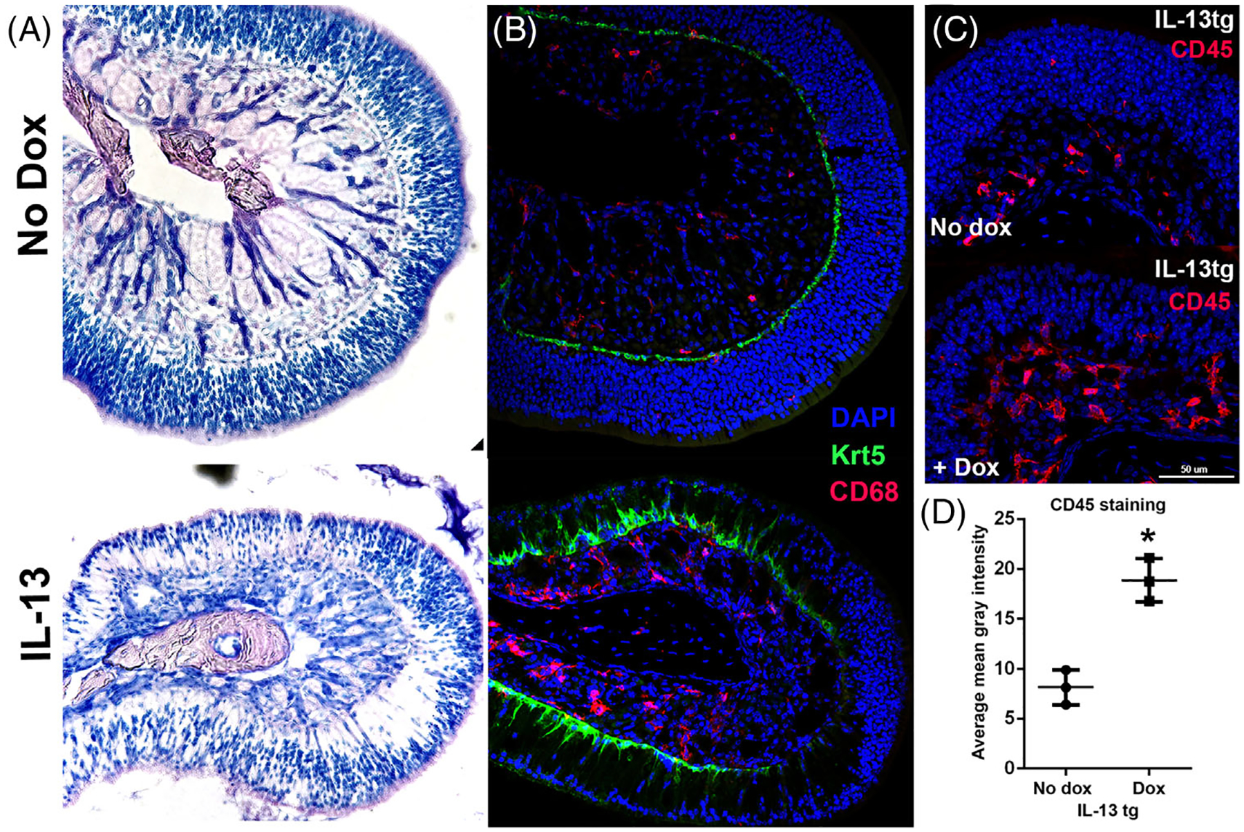
(A) Marked histopathology in the olfactory IL-13 mouse model. Top: normal histology in the absence of doxycycline. Bottom: olfactory turbinate in mouse with IL-13 induction showing regional loss of neurons with maintenance of olfactory thickness. (B) Morphologic change in horizontal basal cells and macrophage infiltrate. Upper image shows normal turbinate in the absence of doxycycline, with flat, quiescent horizontal basal cells (Krt5, green) and few macrophages (CD67, red). Lower image demonstrates pyramidal shape of horizontal basal cells, associated with activation, especially in areas of neuronal loss. Macrophages are prominent in the lamina propria. (C, D) Over twofold increased inflammatory cells in lamina propria in the olfactory IL-13 mice, as quantified by average CD45 staining per tissue area in the lamina propria of turbinate 4E (*p = 0.007). n = 3 mice per group
