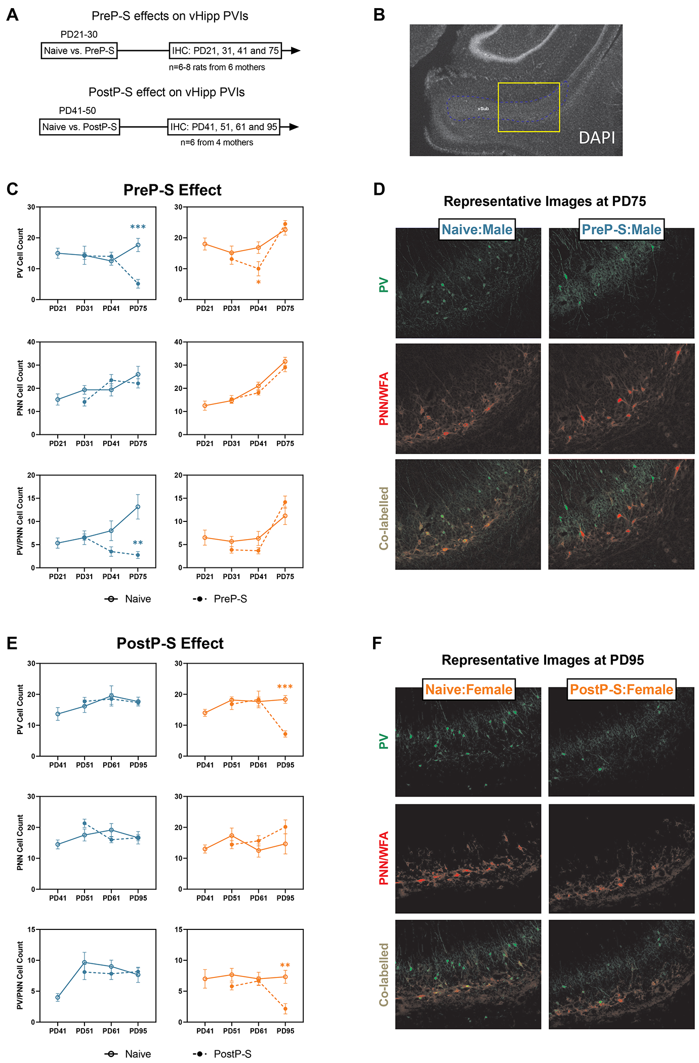
Figure 6. Sex differences in the longitudinal impact of stress on vHipp PV interneurons.

(A) vHipp was sampled at different time points after PreP-S and PostP-S to examine potential stress-induced PV interneuron (PVI) impairments. (B) A 4x image with DAPI staining showing the relative location of the 10x imaging site targeting the proximal portion of the ventral subiculum of the vHipp. (C) PreP-S induced male-dominant PVI impairments in the vHipp. The PreP-S effect in males was delayed, as stress-related reductions of PV+ and PV+/PNN+ co-labeled cell counts were only observed at PD75 (i.e., 45 days after stress). Female vHipp responded to stress with a minor PVI deficit, characterized by a PV reduction at PD41 (i.e., 10 days after stress), which did not persist into PD75. (D) Representative images showing PreP-S effect in males on PVIs measured at PD75. (E) In PostP-S females, stress-related reductions of PV and PV/PNN co-labeled cell count were only observed at PD95 (i.e., 45 days after stress), whereas in males, PostP-S did not induce any change in any examined marker. Data are presented as mean ± SEM. Unpaired t-test with Welch correction was conducted at each post-stress time point. **p<0.01; ***p<0.001, indicating stress-related changes.
