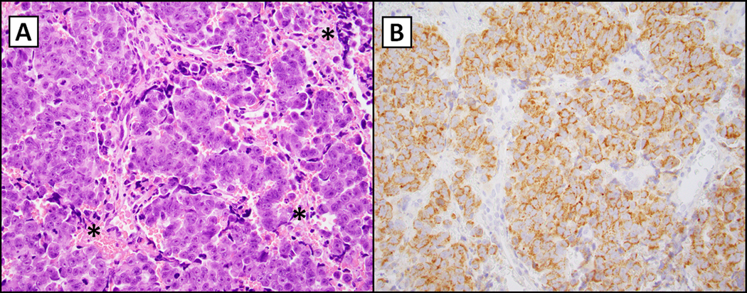
Figure 2.

IDH2-mutated sinonasal carcinoma (R172S; 400X magnification). The left panel (hematoxylin and eosin, A) demonstrates a nested population of epithelioid cells with a prominent nucleolus, variable hyperchromasia and dusky blue cytoplasm typically noted in undifferentiated neoplasms in the head and neck. There is focal necrosis indicated by asterisks. The right panel (B) shows immunohistochemistry for multispecific antibody directed against mIDH1/2 with granular cytoplasmic staining (considered positive if noted in greater than 10% of cells (as seen here)(12). Micrographs courtesy of Dr. Vickie Y. Jo, Brigham and Women’s Hospital, Boston, MA.
