
Figure 7.
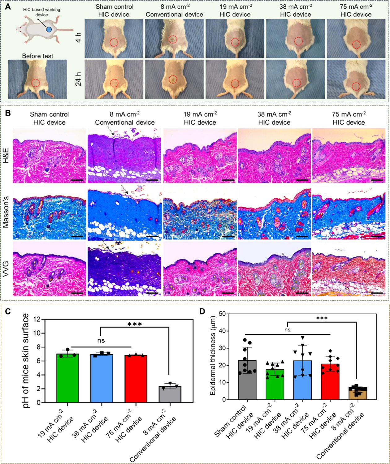
In vivo safety of our high-intensity electrical biofilm treatment system. (A) Representative photographs of mouse in five different treatment groups (0 mA cm−2 (sham control), 19 mAcm−2, 38 mA cm−2, and 75 mA cm−2 1 h treatments applied by our system, and 8 mA cm−2 1 h treatment applied by a conventional electrical device (Figure 2B). Photographs were taken at 4 h and 24 h after treatment. (The upper left schematic was created with BioRender.com) (B) Representative histological sections of skin tissues that were in direct contact with the working device using three stains (hematoxylin and eosin (H&E), Masson’s trichrome, and Verhoeff-Van Gieson (VVG)). Skin samples were collected at 24 h after treatment. Arrows showed the detachment of epidermal from the dermis layer in 8 mA cm−2 conventional device group. Scale bar: 100 μm. (C) Skin surface pH measured immediately after treatment. (D) Thickness of epidermal layer measured from skin sections collected at 24 h after treatment.