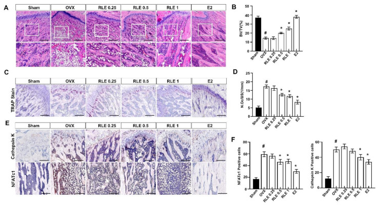
Figure 5.
RLE regulates trabecular bone loss in OVX-induced osteoporosis model. H&E and TRAP staining of distal femoral metaphysis regions. (A) Representative H&E stained femur bone sections (scale bars is 200 μm). (B) The analysis of BV/TV based on the H&E stained femur bone sections. (C) Representative TRAP-stained femur bone. Image J was used to estimate trabecular area and (D) osteoclast number per bone surface. (E) Representative NFATc1 and cathepsin-K-stained femur bone. (F) NFATc1 and cathepsin-K-positive cells were counted and quantified relatively. Data are shown as mean ± SEM. (n = 10, # p < 0.05 vs. sham; * p < 0.05 vs. OVX). Sham, sham-operated group; OVX, ovariectomy; RLE, Ramie leaf extract; TRAP, tartrate-resistant acid phosphatase; N.oc, osteoclast numbers per bone surface.

