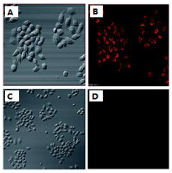
Figure 4.
(A) Bright-field images of cells treated with probe 3. (B) Bright-field images of cells preincubated with probe 3 and NaHS. (B,D) are fluorescence images of (A,C), respectively. Reprinted with permission from Ref. [61]. Copyright ©2017 Royal Society of Chemistry.

