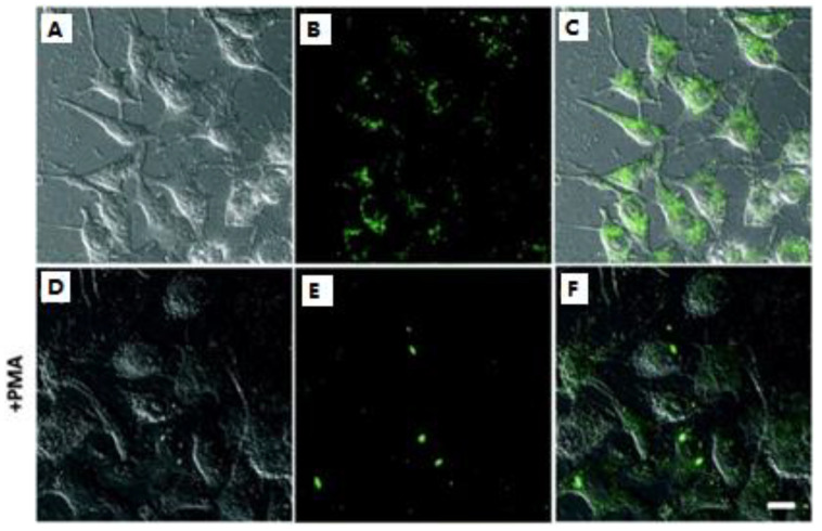
Figure 5.
Fluorescence imaging of the endogenous H2S in the lysosomes: (A–C) the cells incubated with 5.0 μM probe 6 only; (D–F) the cells pretreated with PMA and then incubated with probe 6. (A,D) bright-field images; (B,E) fluorescence; (C,F) merged. Reprinted with permission from Ref. [62]. Copyright ©2016 Royal Society of Chemistry.

