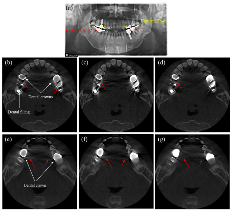
Figure 12.
Images of a patient having multiple dental fillings and dental implants. (a) The panoramic X-ray image, (b,e) the uncorrected CT images, (c,f) the CT images corrected by the DSC method, and (d,g) the CT images corrected by the proposed method. (Contrast window is [−1117~8564] in Hounsfield Unit).

