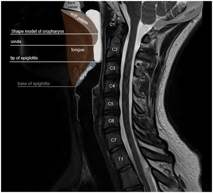
FIGURE 3.

Oropharynx shape model in the context of a T2‐weighted sagittal MRI image. The oropharynx is defined superiorly by the top of the soft palate and inferiorly by the tip of the epiglottis and subdivided into the retropalatal and retroglossal regions posterior to the soft palate and tongue, respectively
