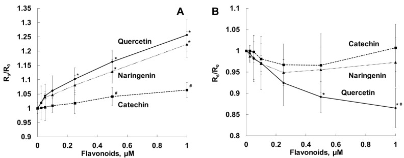
Figure 6.
Dependence of fluorescence anisotropy of DPH, located in the hydrophobic region of the erythrocyte membrane (A), and TMA-DPH, located in the outer monolayer of the erythrocyte membrane (B), on the concentration of the flavonoids. Suspension of erythrocytes (0.01% hematocrit in PBS) was labeled with the fluorescent probes (DPH or TMA-DPH) at a concentration of 1 μM in the dark for 20 min at 37 °C, and then the flavonoids were added and incubated for 30 min at 37 °C. Anisotropy values were registered in the absence (R0) and in the presence (Rs) of the flavonoids. *—p < 0.05 in comparison with dye fluorescence anisotropy in the absence of the flavonoids; #—p < 0.05 in comparison with dye fluorescence anisotropy in the presence of other flavonoids.

