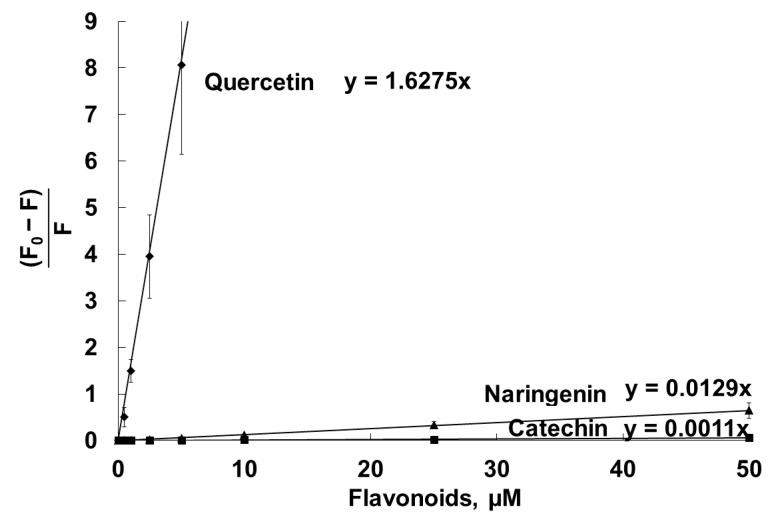
Figure 7.
Stern–Volmer plots of quenching by catechin, naringenin and quercetin of the fluorescence of the DPH probe, located in the hydrophobic part of unilamellar liposomal DMPC vesicles. Liposomes (100 µg/mL, PBS, pH 7.4) were labeled with the DPH fluorescent probe at a final concentration of 1 µM, and then the flavonoids were added and incubated at 25 °C for 30 min. Fluorescence intensity values were registered in the absence (F0) and in the presence (F) of the flavonoids.

