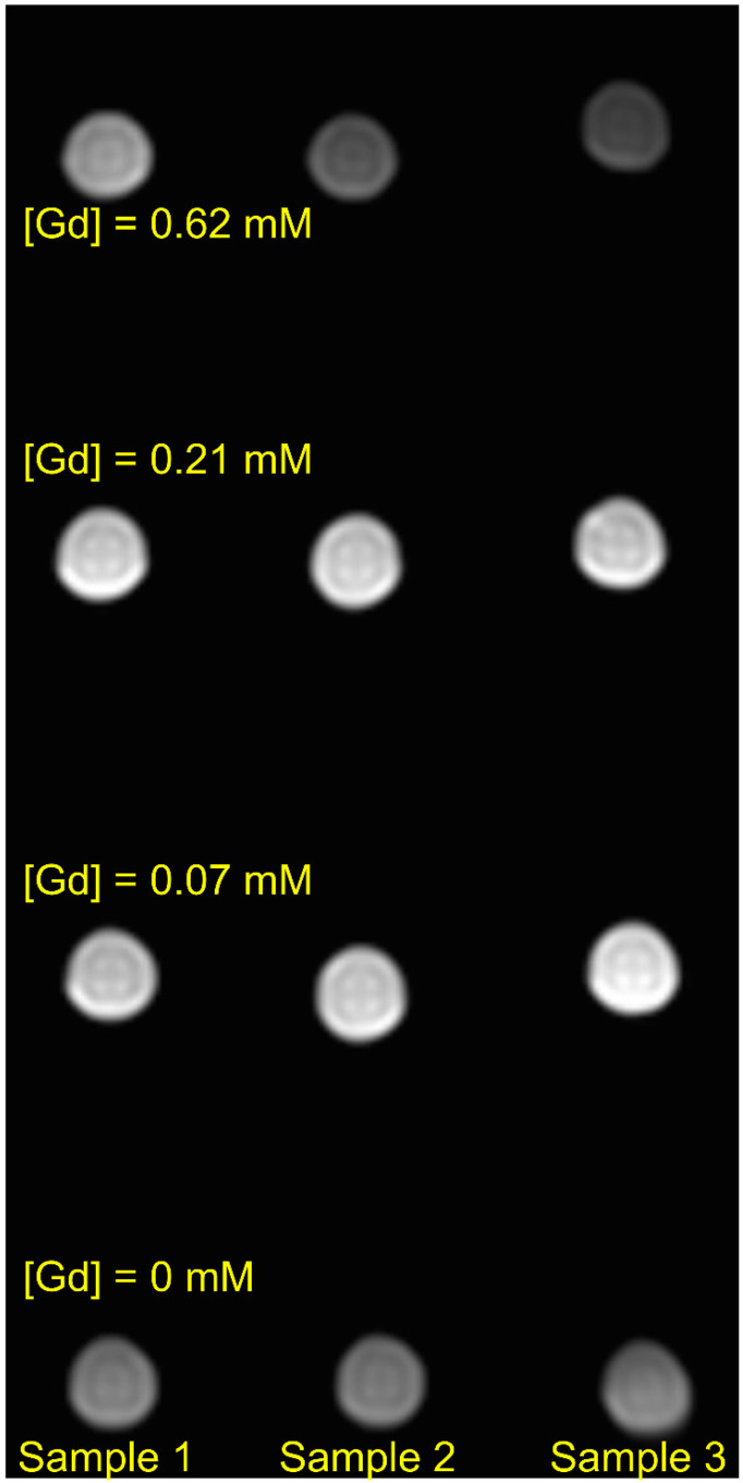
Figure 15.
Combined T1- and T2-weighted image of a cross section of the phantom described in the text. To obtain the image, a T2-TSE pulse sequence was used on a 3 T Philips Ingenia human MRI scanner with parameters ms and ms, (the inversion pulse was not applied). Other parameters of the experiment were: 16-channel head coil for the radiofrequency (RF)-pulse application and detection of proton free induction decay, voxel size 0.5 × 0.5 × 10 mm3, size of image matrix 160 × 160.

