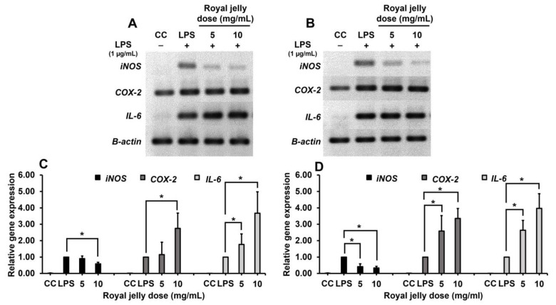
Figure 3.
The inflammatory gene expression of RAW264.7 cells after treatment with royal jelly samples RJ-LP1 (A,C) and RJ-CM1 (B,D) by agarose gel electrophoresis (A,B) and qRT-PCR (C,D). The genes were detected with specific primers (iNOS, COX-2 and IL-6). The data represent mean values of three replicates ± SD. * Royal jelly concentrations were significantly different when compared with LPS control (p < 0.05).

