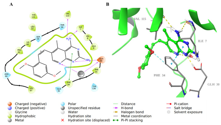
Figure 7.
Molecular docking of co-crystallized inhibitor SRI-9662 in hDHFR (PDB: 1KMV). (A) 2D representation of binding interactions of SRI-9662 with amino acid residues in the active site within a 3 Å distance; (B) 3D representation of SRI-9662 in green within the hDHFR active site. The H-bond and aromatic-hydrogen interactions are in yellow and cyan dotted lines, respectively. A light blue dotted line represents the π-π stacking between aromatic rings.

