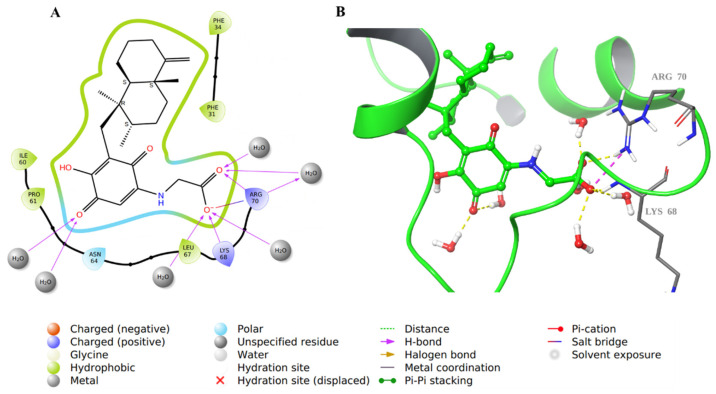
Figure 9.
Molecular docking of co-crystallized inhibitor compound 28 in hDHFR (PDB: 1KMV). (A) 2D representation of binding interactions of 28 with amino acid residues in the active site within a 3 Å distance; (B) 3D representation of 28 in green within the hDHFR active site. The H-bonds and ionic interactions are in yellow and purple dotted lines, respectively.

