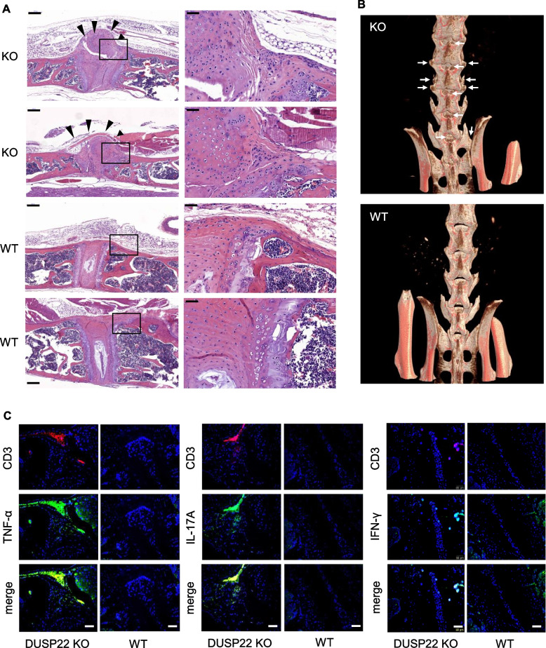
Fig. 5.
Aged DUSP22 knockout mice spontaneously develop AS-like bone disease. A Hematoxylin and eosin-stained sections of vertebral bones from two aged (1-year-old) DUSP22 knockout (KO) mice showed massive mesenchymal cell proliferation and excessive matrix formation bridging across adjacent vertebrae (black arrowheads). Intact joints and intervertebral discs were shown in wild-type (WT) mice. Original magnification, ×10 (left) and × 40 (right). Scale bar, 200 μm (left) and 50 μm (right). B Micro-CT radiographs showed the bone structure of spinal joints, sacroiliac joints, and hip joints of DUSP22 KO and wild-type (WT) mice. Arrows denote osteophytes and bony fusion. C Immunofluorescence assay showed the increase of TNF-α+, IL-17A+, and IFN-γ+ CD3+ T cells in the vertebrae of aged (1-year-old) DUSP22 KO mice. Samples were stained for Alexa Fluor® 488- or PE-conjugated anti-human CD3 antibody, APC-conjugated anti-human TNF-α antibody, FITC-conjugated anti-human IL-17A antibody, and FITC-conjugated anti-human IFN-γ antibody. 4′,6-diamidino-2-phenylindole (DAPI) was used as counterstaining for cell nuclei. Scale bar, 50 μm. One representative image of at least 6 fields is shown

