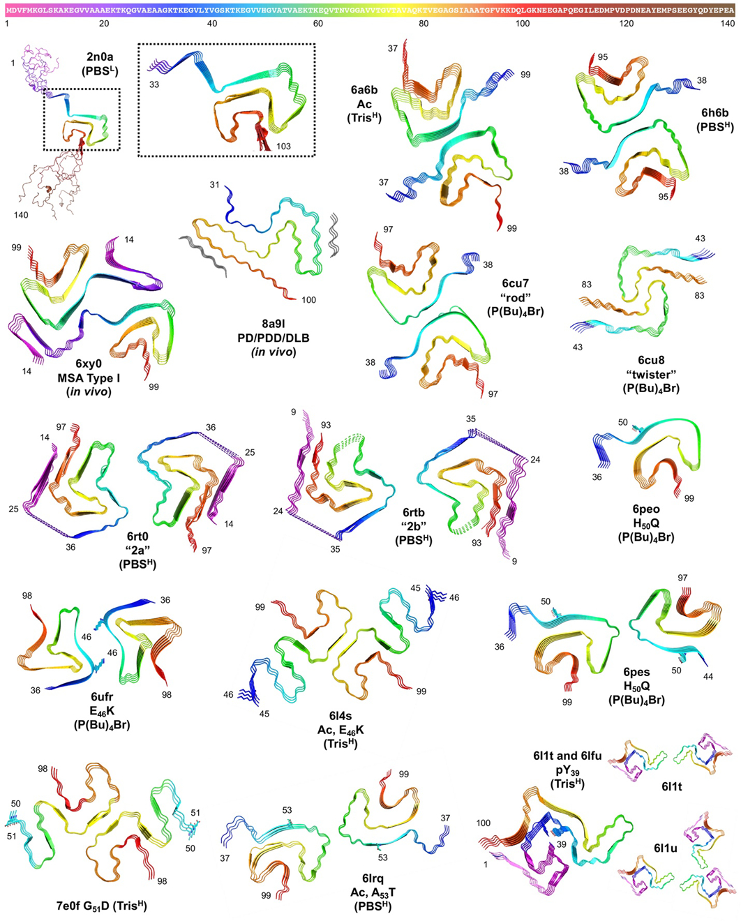
Figure 8.

Top: αS sequence colored as in structures below and Figures 2, 4, 6, and 7. Bottom: Fibril structures from ssNMR (2n0A) and cryo-EM (all other PDB IDs) with αS modifications, aggregation conditions (L = low salt, H = high salt), and key residues labeled and shown as sticks. 6l1t and 6l1u share a common fold with two- or three-stranded fibril packing.
