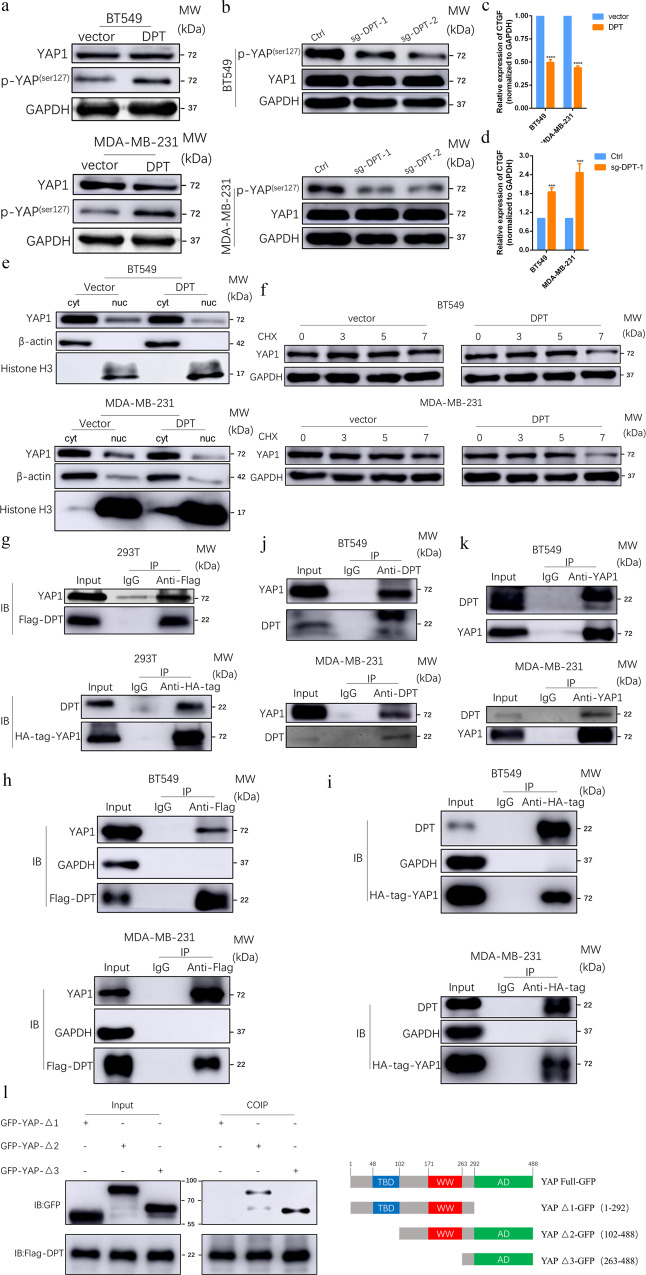
Fig. 5. DPT stably interacts with YAP and suppresses the Hippo/YAP signaling pathway in BC cells.
a Western blotting showed expression level of total YAP and p-YAP in vector and DPT-OE cells. b total YAP and phosphorylated YAP (Ser127) were evaluated by western blot in ctrl and DPT-KO cells. c The mRNA levels of CTGF in DPT overexpression and vector cells. d The mRNA levels of CTGF in ctrl and DPT-KO cells. e Nuclear-cytoplasm separation assay indicated that DPT overexpression regulated YAP translocation between nuclear and cytoplasm. f Protein stability assay indicated the expression of YAP in different time points after Cycloheximide (CHX, 100 μg/ml) treatment in DPT overexpression and vector cells. g–i Immunoprecipitation showed the interaction between exogenous DPT and YAP proteins. j, k Immunoprecipitation assay showed the interaction between endogenous DPT and YAP proteins in BT549 and MDA-MB-231 cells. l Western blot showed that GFP-tagged YAP were precipitated by in vitro transcribed DPT in 293T cells. Right panel: graphic illustration of the YAP domain structure. ***P < 0.001, ****P < 0.0001.

