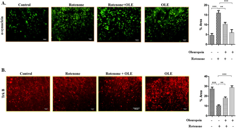
Figure 6.
The immunohistochemical staining of (A) α- synuclein and (B) Trk B. The increase in expression of α-synuclein in rotenone group (***p < 0.001) while neuroprotection of OLE significantly decreased the expression of α-synuclein (**p < 0.01). Similarly, Trk B expression was reduced upon rotenone intoxication (***p < 0.001) whereas, OLE significantly upregulated the expression of Trk B in OLE treated group (**p < 0.01). The one-way ANOVA was used to analyse the data, followed by the Newman–Keuls test. The mean ± SEM (n = 6) is used to depict the values.

