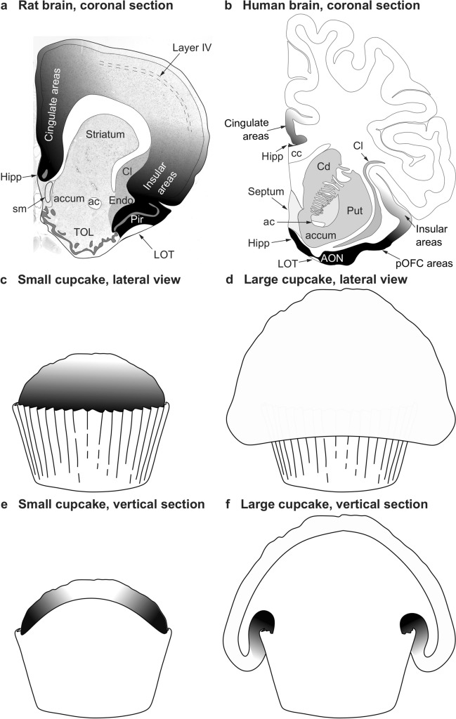
Fig. 1.
Topography and topology of cortical types. a Coronal section of the rat brain through the olfactory tubercule. Dorsolateral isocortical (eulaminate with well-developed layer IV, white and light shades of gray) areas are flanked on the medial side by the allocortical precommissural hippocampus (Hipp, black) and cingulate agranular and dysgranular areas (dark shades of gray); Isocortical areas are flanked on the ventrolateral side by allocortical olfactory areas (Pir, black) and insular agranular and dysgranular areas (dark shades of gray). b Coronal section of the human brain through the nucleus accumbens. Dorsolateral isocortical (eulaminate with well-developed layer IV, white) areas are flanked on the ventromedial side by the allocortical precommissural hippocampus (Hipp, black) and cingulate agranular and dysgranular areas (dark shades of gray); isocortical areas are flanked on the ventral side by allocortical olfactory areas (AON, black) and insular and posterior orbitofrontal (pOFC) agranular and dysgranular areas (dark shades of gray). c Lateral view of a small cupcake. d Lateral view of a large cupcake. e, Vertical section of the small cupcake in c; the gray scale in c and e resembles the distribution of cortical types on the rat brain in a. f Vertical section of the large cupcake in d; the gray scale in d and f resembles the distribution of cortical types on the human brain in b. The topological neighborhood relations of cortical types (shades of gray) in a, c, and e are preserved in b, d, and f, despite the extensive expansion of isocortical areas in primates that expand over limbic areas like cupcake batter flows over a cupcake mold. ac anterior commissure, accum nucleus accumbens, AON anterior olfactory nucleus in the primary olfactory cortex, cc corpus callosum, Cd caudate nucleus, Cl claustrum, Endo endopiriform nucleus, Hipp anterior extension of the hippocampal formation, LOT lateral olfactory tract, Pir piriform cortex in the primary olfactory cortex, pOFC posterior orbitofrontal cortex, Put putamen, sm stria medullaris, TOL olfactory tubercule. a, b are modified from Fig. 3 in Garcia-Cabezas and Zikopoulos (2019)

