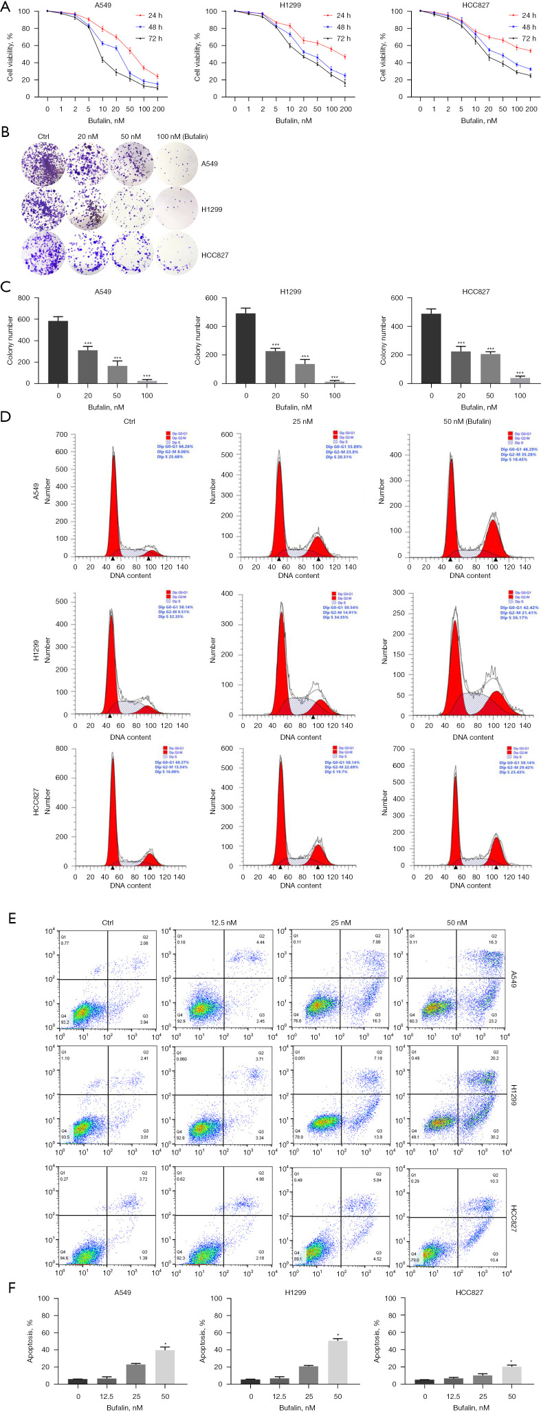
Figure 1.
Bufalin decreased cell growth and viability in NSCLC. (A) Three NSCLC (A549, H1299, and HCC827) cell lines were incubated with bufalin at various concentrations (0 to 200 nM) for 24, 48, and 72 h, and then, the cell viability was assayed by CCK-8 assay. (B,C) Representative images of colonies stained with crystal violet after treatment with DMSO, 20, 50, and 100 nM bufalin. The colony numbers are shown on the right (magnification, ×1). (D-F) After treating cells with different concentrations of bufalin for 48 hours, the cell cycle and apoptosis were examined by flow cytometry. The apoptosis rates are shown on the right. *, P<0.05; ***, P<0.01 vs. untreated controls. Ctrl, control; NSCLC, non-small cell lung cancer; CCK-8, Cell Counting Kit-8; DMSO, dimethyl sulfoxide.

